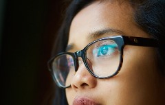
May Is Vision Health Month
Keep your family’s eye healthy
Vision loss can happen to anyone, at any age. In fact, one in seven Canadians will develop a serious eye disease in their lifetime. But did you know that 75 per cent of vision loss can be prevented or treated? Preventive measures and early detection of eye disease significantly lowers your risk of vision loss.
Each year, we celebrate Vision Health Month in May.
Vision Health Month is a national awareness campaign designed to educate Canadians about their vision health and eliminate avoidable sight loss across the country. Throughout the month of May, CNIB asks all Canadians to protect their families’ vision health, starting with getting regular eye exams from a Doctor of Optometry.
Prepare an eye-healthy meal
Did you know that nutrition can have a profound influence on your vision health? Good nutrition for the eyes means a balance of vitamins, minerals, fats and other nutrients. But negotiating your way through a maze of nutrient-dense lingo can be difficult. Check out our eye-healthy recipes for an easy way to protect your family’s sight!
Seeing Beyond Vision Loss
Every 12 minutes, someone in Canada begins to lose their eyesight. But did you know that 75 per cent of vision loss can be prevented?
In this section, you’ll find valuable tips to help you protect your eyes from injury, prevent vision loss before it starts, or treat vision loss that may have already begun. You’ll also find clear information about any eye condition you may have, and expert answers to common questions about vision health and vision loss.
Eye Conditions
Vision loss can be caused by eye problems that are present from birth, by conditions that appear later in life, or by infections or environmental factors.
In this section, you’ll find information about some common eye problems as well as the main causes of vision loss in children and adults.
AGING EYES
As people age, they often begin to have difficulty focusing their eyes for reading or close work. This is called presbyopia and is the normal aging of the eyes. It usually affects people over the age of forty as the eye starts to lose some of its flexibility.
Treatment
This condition is easily corrected with eye glasses of increasing strength as a person ages. Bifocal of trifocal lenses may be prescribed to some people with presbyopia who also have other refractive problems such as myopia (near-sightednness), hyperopia (far-sightedness) or astigmatism (distorted vision due to an irregularly shaped cornea).
AMB (AGE-RELATED MACULAR DEGENERATION)
Age-related macular degeneration (AMD) is a common eye disease, affecting one million people in Canada. More Canadians have AMD than breast cancer, prostate cancer, Parkinson’s or Alzheimer’s combined. Yet few Canadians even realize it exists.
AMD is a progressive eye disease that affects central vision. People who have AMD may no longer be able to read, drive or see the faces of their family members. For many people, the personal, social and economic costs of AMD can be extremely challenging. And with an aging population, the number of people with the disease is expected to double in the next 25 years.
Without treatment, AMD can advance to uncorrectable vision loss. But with proper management, regular eye exams and help from the members of your health care team, you can slow down or even stop the progression of vision loss, and learn some helpful tips that will help you fully participate in life, despite vision loss.
ASTIGMATISM
In normal, undistorted vision, the cornea (the clear window in front of the eye) is smooth and equally curved in all directions. With astigmatism, the cornea is “warped”, meaning it curves more in one direction than the other distorting or blurring vision for objects at any distance.
Large amounts of astigmatism are usually inherited, present at birth and frequently remain unchanged throughout life. Small amounts of astigmatism can be acquired any time in life and are, in fact, very common. It often does not require correction.
Treatment
Correction is not difficult if the distortion proceeds across the cornea in a regular direction. Prescription glasses can often be ordered that neutralize or off set the distortion to the cornea.
If, however, the distortion is irregular, only reshaping the cornea will correct the problem. This is usually done through the use of hard lenses or by replacing it with donated human corneal tissue. While hard lenses are usually better than soft lenses for this correction, some special soft lenses may be helpful to correct mild astigmatism. Large amounts of astigmatism are not easily corrected with lenses as it may wobble on the uneven surface of the cornea. In such instances, a special device called a toric lens may be ground with a curve on the back surface which stabilizes the lens on the cornea.
CATARACTS
More than 2.5 million Canadians have cataracts. They are painless, develop within the existing lenses in your eyes, and are usually detected during routine eye exams.
As you age, your lenses naturally harden and may also turn cloudy. A cloudy lens blocks light from reaching the retina and interferes with vision – the effect is similar to looking through a dirty car windshield. Cataracts may form in one or both eyes, at the same time or at different times.
Fortunately, cataracts can be removed and your vision can be restored. Surgery is usually recommended once a person feels their vision interferes with daily activities such as driving, reading or watching TV.
Cataract surgery is among the most highly perfected, safe and successful procedures in medicine – more than 95 per cent of patients have improved vision after surgery. It removes the cloudy lens and replaces it with an artificial one.
Symptoms + Risk Factors
· Sensitivity to bright light or experiencing glares and haloes around lights
· Difficulty seeing details and poor central vision
· An inability to distinguish colours
· Difficulty seeing at night
· Double vision
Risk Factors:
· Age
· Family History
· Other health problems – complications such as diabetes
· Smoking
· Drinking alcohol
· Excessive sun exposure
· Medications
· Eye injury (puncture, cut, intense heat or chemical burn to the eye)
CHARLES BONNET SYNDROME
Charles Bonnet Syndrome (CBS) is a common condition among people with serious vision loss characterized by temporary visual hallucinations.
Charles Bonnet Syndrome is not a mental illness, nor is it a symptom of dementia or any other disease. Rather, it is a condition specifically related to vision loss. While it is actually very common, awareness is limited, which can cause confusion and concern among those experiencing and diagnosing it.
While it is not yet known exactly why these hallucinations occur, researchers are beginning to believe they are related to an attempt by the brain to fill in information that would normally be obtained from the eyes.
Symptoms
CBS hallucinations tend to begin in the months immediately following a loss of sight. Hallucinations can be very distressing, but aren’t usually permanent. Symptoms generally decrease in frequency after about a year to 18 months, although some people may have the condition for five years or more.
Images can appear at any time and can last from just a few seconds to several minutes.
People with CBS hallucinations are usually aware that what they’re seeing is not real. CBS hallucinations only affect sight, which means that people with CBS don’t hear, smell or feel things that aren’t there.
These visual hallucinations can take many forms, ranging from simple shapes and lines to detailed images of people and landscapes. They may seem “real” (like seeing dragons). Most frequently, people will see patterns or simple shapes, however there are many accounts of more complex hallucinations,
including:
· Little men holding umbrellas at the end of the bed
· Women in red dresses sweeping the floor
· Cloakroom tickets lining the walls and ceiling
· Soldiers marching down the street
It’s clear that CBS affects every person differently, so it’s important to choose a coping strategy that works for each unique case.
DIABETIC RETINOPATHY
Diabetes can affect many parts of the body, and people with diabetes are at a high risk of developing vision problems, usually from a condition called diabetic retinopathy. It’s estimated that half a million Canadians have diabetic retinopathy.
Without treatment, the condition can lead to uncorrectable vision loss or even blindness, usually in both eyes. In fact, diabetes is the leading cause of blindness in Canadians under 50. But with regular comprehensive eye exams by an eye doctor, diabetic retinopathy can be detected and treated, often prolonging the ability to see and preventing further damage.
DIABETIC MACULAR EDEMA
Diabetic macular edema (DME) is a common complication of diabetic retinopathy (DR). DME is caused by damage to the blood vessels of the retina and is a leading cause of vision loss in the working-age population in the developed world.
When the blood vessels in the retina swell and leak, the leaking fluid can cause swelling in the macula, the central part of the retina at the back of the eye where sharp vision is determined. Macular edema – the swelling of the macula – can then result in blurred vision and can lead to permanent vision loss over time.
While DME can occur at any stage of diabetic retinopathy, DME is more likely to occur as the disease progresses.
It is estimated that vision impairment resulting from DME affects approximately 2.5% of the nearly 2.4 million Canadians with diabetes, or approximately 60,000 Canadians, making it a major cause of adult-onset vision loss.
DRY EYE
Simply put, dry eye is the result of not being able to produce enough tears to keep the eye comfortable. Without a film of tears, spread over the eye by a blink, good vision is not possible.
The symptoms include stinging or burning eyes, scratchiness, stringy mucus around the eyes, irritation from smoke or wind, excess tearing or difficulty wearing contact lenses.
Causes
Tear production normally decreases as we age. Although dry eye can occur in men and women, it is more common in women, particularly after menopause. Dry eye can also be associated with arthritis as well as a variety of over-the-counter and prescription medications. Because of this—and the fact that people with dry eye are more prone to the toxic side effects of eye medications—it is important that you tell your doctor the names of all the medications you are taking. This is especially true if you are taking diuretics, betablockers, antihistamines, sleeping pills, pain relievers, medication for “nerves” or even alcohol.
FAR-SIGHTEDNESS (HYPEROPIA)
Hyperopia (far-sightedness), myopia (near-sightedness), and astigmatism (distorted vision) are what known as refractive errors.
For proper eyesight, the cornea (the clear window I front of the eye) and the lens (behind the pupil) must properly focus or “refract” light onto the retina (at the back of the eye). If the length or shape of the eye is not idea, the light may get focused too early or too late leaving a blurred image on the retina.
Hyperopia, or far-sightedness, is the ability to clearly see objects at a distance but not those up close.
Causes and Symptoms
Like near-sightedness, far-sightedness is usually an inherited condition. Young children tend to be hyperopic to some degree although the severity lessens as they age and the eye grows and becomes longer.
Children with far-sightedness can often see both distant and close objects because the youthful strength of their lenses can often overcome the shortness of the eye. Non-visual signs such as headaches or a lack of interest in reading may be signs of a high degree of hyperopia.
FLOATERS AND FLASHING LIGHTS
Floaters are dark specks in the form of dots, circles, lines, or cobwebs that seem to move across your field of vision. They are most noticeable when you are looking at a light-coloured background, such as a clear sky or a white wall. Floaters come in many sizes and numbers, and they seem to move when you look in different directions.
What causes floaters?
Floaters are dark specks in the form of dots, circles, lines, or cobwebs that seem to move across your field of vision. They are most noticeable when you are looking at a light-coloured background, such as a clear sky or a white wall. Floaters come in many sizes and numbers, and they seem to move when you look in different directions.
Floaters are small clumps of visible material within the vitreous. Although floaters seem to be in front of you, they are actually floating within the vitreous and the retina sees them as shadows. Sometimes floaters occur because your eye is irritated, or because there are crystal-like deposits in the vitreous. As we get older, the vitreous shrinks and separates from the retina. As this separation occurs, floaters may appear. In about half of all people, the vitreous has separated from the retina by age 50, although many people are not aware that this process has occurred in their eye(s).
Most of the time, the vitreous separates from the retina without any problem. However in some people, the vitreous remains attached to parts of the retina, causing the retina to tear as the vitreous separates from it. There may be some bleeding into the vitreous associated with a retinal tear, which results in a new shower of floaters.
Tears in the retina should be repaired right away. A retinal tear can develop into a retinal detachment, which is a serious threat to vision. Retinal tears can usually be repaired without a hospital stay, but it may take a major operation to fix a retinal detachment.
Certain people, such as those who are nearsighted and those who have had a cataract operation or an eye injury, have a higher risk of retinal tears and retinal detachment.
What are flashing lights?
The vitreous sometimes pulls on the retina. This may cause you to see flashing lights or lightning streaks, even though there aren’t any such lights present. This is the same thing that occurs when you get hit in the eye and “see stars” or when you rub your eye. When the vitreous separates from the retina, you may see flashing lights once in a while, for a few weeks.
If you see jagged lines of light that last 10 to 20 minutes and happen in one or both eyes, this is probably a migraine caused by the spasm of blood vessels in the brain. If you develop a headache after, it is called a “migraine headache.” If you don’t develop a headache after, the light flashes are called “ophthalmic migraine,” or “migraine without headache.
GLAUCOMA
Glaucoma is the second most common cause of vision loss in seniors in Canada. More than 250,000 Canadians have chronic open-angle glaucoma, the most common form of the disease.
Glaucoma involves damage to the optic nerve most often caused by high pressure inside the eye due to a build-up of excess fluid. High eye pressure is not always a sign that you have glaucoma but may be an indication you are at risk of developing it.
Over time the disease develops into a loss of peripheral (side) vision. If glaucoma is untreated it could advance to later stages where central vision narrows to “tunnel” vision, or it may result in complete loss of vision. Early detection and treatment is essential to prevent severe vision loss or blindness.
Types of Glaucoma + Risk Factors
Primary Glaucoma
Open-angle glaucoma accounts for 90 per cent of all cases in Canada – Initially you can perform all normal daily activities such as driving and reading and vision loss is not obvious until it is too late and permanent.
Primary acute closed-angle glaucoma results from a buildup of fluid in the eye because the distance between the iris and the drainage system has been closed, stopping fluid from draining from the eye. Occurs very suddenly and is an emergency.
Secondary Glaucoma can result from a variety of other conditions such as an eye injury or inflammation, eye surgery complications, diabetes and the use of certain medications.
Risk Factors:
· Elevated pressure in the eye
· Family history
· Age – after age 40 and after age 60
· Ethnicity
· Myopia
· Diabetes
LAZY EYE (AMBLYOPIA)
Amblyopia (‘lazy eye’) happens when the vision in one eye doesn’t develop properly in early childhood. You may not be able to notice it easily in your child, but if it isn’t treated it will become a permanent visual problem.
Babies are able to see when they are born, but they have to learn how to use their eyes. They have to learn how to focus, and then how to use both eyes together. Our vision continues to develop until we are about 9 to 12 years old. After that, our eyesight is complete and can’t be easily changed.
However, sometimes the vision in one eye doesn’t develop properly. This may be caused by misaligned eyes (called strabismus) or because one eye is out of focus compared with the other. When this happens, the brain ‘shuts off’ the eye that is out of focus, and the child depends only on the better eye to see.
An eye disease such as a cataract or anything else that stops a clear image from being focused inside the eye can cause amblyopia in children. They may also inherit conditions from their parents that lead to amblyopia.
NEAR-SIGHTEDNESS (MYOPIA)
Myopia (near-sightedness), hyperopia (far-sightedness), and astigmatism (distorted vision) are what as known as refractive errors.
For proper eyesight, the cornea (the clear window in front of the eye) and the lens (behind the pupil) must properly focus or “refract” light onto the retina (at the back of the eye). If the length or shape of the eye is not ideal, the light may get focused too early or too late leaving a blurred image on the retina.
Myopia, or near-sightedness, is the ability to clearly see objects up close but not those at a distance.
Causes
It is an inherited condition usually detected in children between the ages of eight and twelve. Few factors outside of heredity affect this condition. Using dim light, reading too much or nutritional deficiencies do not seem to impact it one way or the other.
PINK EYE (CONJUNCTIVITIS)
Conjunctivitis, better known as pink eye, is the medical term that describes an inflammation of the conjunctiva, the thin membrane that covers the white of your eyes.
This membrane produces mucus to coat and lubricate the surface of the eye. Upon close inspection, you can see fine blood vessels within this membrane. When the conjunctiva becomes irritated, the blood vessels enlarge making the eye appear red in colour.
Causes and Symptoms
Conjunctivitis can be caused by a number of things, but the most common are viral and bacterial infections, allergies or environmental irritants. Infection conjunctivitis—whether viral or bacterial—can be quite contagious, so contact with the infected person’s tears through handkerchiefs and towels should be avoided. Hand washing after contact will also prevent the spread.
The signs are usually quite obvious: red, irritated eye; a burning or scratchy feeling; or, a pus-like discharge.
There are several eye diseases which also produce what might be confused with “pink eye”, so a medical evaluation with an ophthalmologist—a medically trained eye specialist—is always a good idea. This is particularly true if pain, blurred vision or severe light sensitivity are also present as these are not normally associated with conjunctivitis.
RETINAL DETACHMENT
Retinal detachment is a serious problem that usually affects middle-aged or older people. If it isn’t treated immediately, it will lead to vision problems or even vision loss.
In most cases the retina detaches because of small tears or holes. These may occur as the retina thins with age. More often, they happen because the vitreous partially separates from the retina. The vitreous is firmly attached to the retina in a few places, and as we get older it separates and may tear the retina at those areas of attachment. In about half of the population, the vitreous has separated from the retina by age 50.
Nearsightedness, infection or injury such as a hard solid blow to the eye, can also make the vitreous separate. It is more common in people with a family history of retinal detachment. When there is a hole in the retina, fluid from the vitreous space may pass through the hole and flow between the retina and the back wall of the eye. This can separate the retina from the back of the eye and cause it to detach. The detached part of the retina will not work properly, and you will see a blurry or blind spot in your field of vision.
Less commonly, retinal detachment can also be caused by eye diseases, complications of diabetes, or tumours. In these cases there may be no tears or holes in the retina.
RETINITIS PIGMENTOSA
Retinitis pigmentosa (RP) refers to a group of diseases which tend to run in families and cause slow, but progressive loss of vision. The retina is the tissue which lines the inside of the eye and sends visual images to the brain. In RP, there is gradual destruction of some of the light sensing cells in the retina.
Causes and Symptoms
The cuase of retinitis pigmentosa has not been clearly identified. About fifty percent of RP cases are sporadic with the remaining cases being hereditary. Learning more about the history of people with RP may help predict how the disease will progress for the affected person, although variations can exist even from person to person within a family.
The first symptoms usually start during young adulthood, although it may be seen first at any age. The two most common symptoms are night blindness (where adjusting to the dark happens very slowly) and the loss of side vision. Loss of side or peripheral vision makes mobility very difficult. Once this occurs, a person is left with only central vision (as if looking down a tunnel).
RETINAL VEIN OCCLUSION
Like many Canadians, you may have never heard of Retinal Vein Occlusion (RVO) or macular edema. RVO is a disorder of the retina that can lead to vision loss and cause blindness in some patients.
RVO is a blockage of the veins in the retina that carry blood away from the eye. The blockage can cause fluid to leak into the macula – the part of the eye where focusing occurs. The swelling, called macular edema, can cause blurred vision and sometimes complete vision loss.
The blockage caused by RVO cannot be reversed, but there are ways in which it can be managed, using treatments that may slow down the progression of vision loss and even greatly improve your vision.
TURNED EYES (STRABISMUS)
Strabismus is the medical term for misalignment of the eyes, two eyes that are not straight. It occurs in at least 5% of the population. There are three common types:
1. Esotropia, or ‘crossed eye’ – One eye is turned inward, toward the nose a child may be born with this condition, or it may develop a few months after birth or around age two to three.
2. Exotropia, often called ‘wall-eye’ in the past – One eye is turned outward, away from the nose. A child is rarely born with this condition. More often, it develops later.
3. Vertical strabismus – The eyes are out of alignment vertically.
Strabismus can cause of couple of different vision problems:
· Defective binocular vision: the eyes need to be straight for the brain to combine the images seen by the two eyes into a single picture. This gives us 3-D vision, which allows us to judge depth. Any turn of the eye can interrupt 3-D vision. As well, if an eye turns in, it can reduce the total field of vision.
· Vision loss in the turned eye: When a child’s eye is turned, he or she may end up with only partial sight in the turned eye. This condition is called amblyopia, or ‘lazy eye.’ This can also happen if the eyes are unequally focused.
Click here to learn more about vision health
Source: cnib.ca